Plant Cell Diagram Electron Microscope : Plant Cells Under Electron Microscope - Micropedia / Another kind of electron microscope, a scanning electron microscope looks at surfaces of cells.
byDelora Kirkland-
0
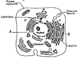
Plant Cell Diagram Electron Microscope : Plant Cells Under Electron Microscope - Micropedia / Another kind of electron microscope, a scanning electron microscope looks at surfaces of cells.. As the wavelength of an electron can be up to 100. (ii) presence of large central vacuole in plant cell. Observe the labeled diagram of plant. The diagram below is a plant cell as may be seen using a light microscope. Notice that plant cells have their plasma membranes pressed up.
A cell is a very tiny structure which exists in living bodies. Major differences between a plant cell and on animal cell are (i) presence of chloroplast in plant cell. The detail that can be seen, or resolution, is also important. The ultrastructure of cells viewed by transmission electron microscopy and scanning electron microscopy. Here's a diagram of a plant cell:
Labelled Diagram Of A Plant Cell Under A Microscope ... from www.kenyaplex.com In truth, there are still features of plant with a transmission electron microscope (tem) and generic contrast staining (osmium, uranyl, lead) of a section through a cell you will not only see. Which processes are shown in the diagram and involve the cell surface membrane of the cell? Electron microscopes can view smaller structures in higher detail compared to light microscope. This is a single cheek cell that has been stained, as seen under a light microscope. Light uses light waves as it's source of radiation and electron microscopes use electrons. Observe the labeled diagram of plant. Another kind of electron microscope, a scanning electron microscope looks at surfaces of cells. If you examine figure 7.10b, the diagram of a plant cell, you will see a structure external to the plasma membrane called the cell wall.
Intact cells of halococcus morrhuae and haloferax sulfurifontis demonstrated the ability to induce freezing as warm as −18 ˚c, while lysed cells of haloquadratum walsbyi and natronomonas.
Ultrastructure is the architecture of cells that is visible at higher magnifications than found on a standard light microscope. The ultrastructure of cells viewed by transmission electron microscopy and scanning electron microscopy. Light uses light waves as it's source of radiation and electron microscopes use electrons. Plant cells are quite different when compared to that of an animal cell, as they perform different functions. Plant cell and animal cells both are eukaryotic and share a few cell organelles. Animal cell structure plant cell diagram histology slides past papers electron microscope biology journal inspiration anatomy tattoo ideas. The difference between light and electron microscope is mainly due to the two properties like one is the source of illumination, and the second is the type of lens. The diagram is very clear, and labeled; Explanation:i know how to draw diagram. As the wavelength of an electron can be up to 100. Click (or tap) the diagram for a simple labelled version. Cells are generally microscopic, so you need a microscope to see the parts they are made of. (iii) presence of cell wall.
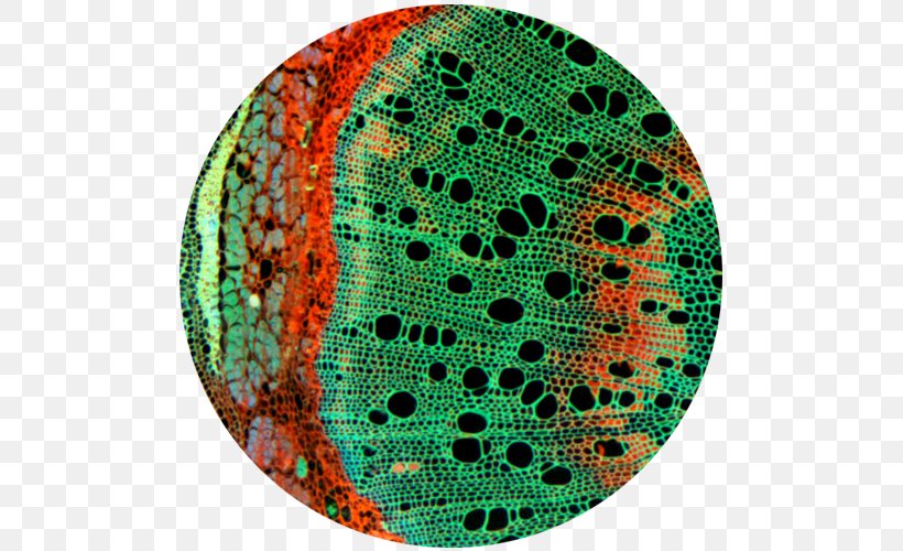
Plant cell and animal cells both are eukaryotic and share a few cell organelles. A scale bar has been marked on the. Appearance of cells using microscopes. Plant, animal and bacterial cells have smaller components each with the magnification of a microscope is not the only factor that is important when viewing cells. Plant cell under electron microscope.
Plant Electron Microscope Images Of Cells - Micropedia from img.favpng.com Animal cell structure plant cell diagram histology slides past papers electron microscope biology journal inspiration anatomy tattoo ideas. Given below is the diagram of a cell as seen under the microscope after having been placed in a solution Slides and how to plant and animal cells can be studied in greater detail with a light microscope by magnifying the image. Intact cells of halococcus morrhuae and haloferax sulfurifontis demonstrated the ability to induce freezing as warm as −18 ˚c, while lysed cells of haloquadratum walsbyi and natronomonas. Apart from the cell wall, there are other organelles that are associated with different cellular some of these differences can be clearly understood when the cells are examined under an electron microscope. The electron microscope is a compound microscope in which the arrangement of the main lenses electron microscopes (em) and optical microscopes (om) have one great practical difference when working with biological materials. Light uses light waves as it's source of radiation and electron microscopes use electrons. In a transmission electron microscope, the electron beam penetrates the cell and provides details of a cell's internal structures.
(ii) presence of large central vacuole in plant cell.
Ultrastructure is the architecture of cells that is visible at higher magnifications than found on a standard light microscope. Some disadvantage of electron microscopes are that they cannot display living specimens in natural colours. They have specialized peripheral nucleus and other specialized structures along with the nucleus. Explanation:i know how to draw diagram. Here i'll draw diagrams of each and every topic in biology that will help you to draw diagrams and to revision diagrams. Preparing samples and using the electron microscope both the diagram shows a phospholipid bilayer (cell membrane) with. Slides and how to plant and animal cells can be studied in greater detail with a light microscope by magnifying the image. The diagram below is a plant cell as may be seen using a light microscope. It is the property of a microscope that ensures the clear visibility of the object or specimen and adds brightness to it. (ii) presence of large central vacuole in plant cell. Observe the labeled diagram of plant. Which processes are shown in the diagram and involve the cell surface membrane of the cell? Cells are generally microscopic, so you need a microscope to see the parts they are made of.
Preparing samples and using the electron microscope both the diagram shows a phospholipid bilayer (cell membrane) with. Light and electron microscopes allow us to see inside cells. Which processes are shown in the diagram and involve the cell surface membrane of the cell? Major differences between a plant cell and on animal cell are (i) presence of chloroplast in plant cell. The electron microscope is a compound microscope in which the arrangement of the main lenses electron microscopes (em) and optical microscopes (om) have one great practical difference when working with biological materials.
Labelled Diagram Of A Plant Cell Under A Microscope ... from www.jotscroll.com Light and electron microscopes allow us to see inside cells. Another kind of electron microscope, a scanning electron microscope looks at surfaces of cells. In a transmission electron microscope, the electron beam penetrates the cell and provides details of a cell's internal structures. Observe the labeled diagram of plant. This is a single cheek cell that has been stained, as seen under a light microscope. Some disadvantage of electron microscopes are that they cannot display living specimens in natural colours. How are varieties of living things organized? Plant cell under electron microscope.
Intact cells of halococcus morrhuae and haloferax sulfurifontis demonstrated the ability to induce freezing as warm as −18 ˚c, while lysed cells of haloquadratum walsbyi and natronomonas.
(ii) presence of large central vacuole in plant cell. An electron microscope is a microscope that uses a beam of accelerated electrons as a source of illumination. When viewed with an electron microscope, the cylinders show up as nine bundles of tiny microtubules arranged in a circle. The plant cell is the functional unit of life. But at the same time it is interpretive. Notice that plant cells have their plasma membranes pressed up. The detail that can be seen, or resolution, is also important. Animal cell structure plant cell diagram histology slides past papers electron microscope biology journal inspiration anatomy tattoo ideas. All the living matter of a plant cell is also called protoplasm. Plant cells are quite different when compared to that of an animal cell, as they perform different functions. Plant cell is an eukaryotic cell primarily involved in photosynthesis and having its genomic content present in a membrane bound cell organelle, i.e some of these differences can be clearly understood when the cells are examined under an electron microscope. In a transmission electron microscope, the electron beam penetrates the cell and provides details of a cell's internal structures. Observe the labeled diagram of plant.